Our research combines experimental imaging—including MRI pulse sequence development—and computational and analytical signal modelling to develop ways to image brain activity and physiology. Modelling helps to guide our experiments, and experiments provide data for our modelling. Imaging research is conducted on a research-dedicated, hybrid Siemens 3-T PET-MRI scanner located at the Brain Imaging Centre at The Royal. While most of our research is conducted on healthy volunteers, our lab also has collaborations with scientists at the IMHR to provide more quantitative measurements of brain physiology in mental illness, including bipolar disorder and treatment-resistant depression.
Physiological imaging
The main thrust of our research is the development of techniques that quantitatively image brain physiology using MRI. We use and optimize a range of imaging contrasts to measure specific parameters of interest relevant for mapping brain activity and overall brain health, including cerebral blood flow (CBF) and volume (CBV), the cerebral metabolic rate of oxygen (CMRO2), blood oxygenation, and the oxygen extraction fraction (OEF). We measure these parameters both at rest and dynamically during tasks that induce changes in brain activity or by having participants breathe from a gas delivery system that supplies air with elevated levels of carbon dioxide, to temporarily change blood flow, or oxygen, to change blood oxygenation.
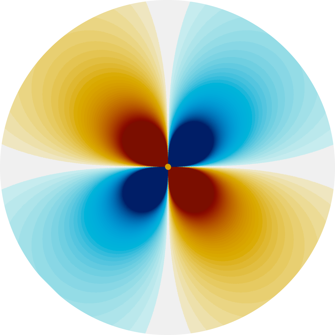
Biophysical modelling
Biophysical modelling is a key tool in the development of new imaging techniques and it has been invaluable in informing our interpretation of most fMRI studies. Biophysical models allow us to capture various aspects of brain physiology, e.g., the vasculature and oxygenation of blood, and determine how they impact the measured MRI signal. We develop stochastic (i.e., Monte Carlo methods) and deterministic simulation methods and a range of geometries to represent the vasculature and red blood cells. These variations in simulation approaches allow us to trade off model complexity, accuracy, and computational demands. Much of this modelling work is implemented in and enabled by BOLDsωimsuite, an open-source Python toolkit that we develop in collaboration with scientists at the University of Waterloo and the University of Toronto.
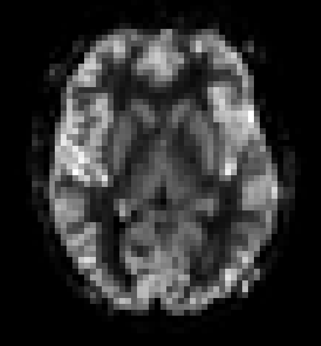
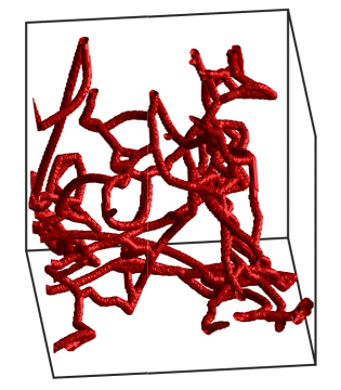
High-resolution imaging
We seek to improve the anatomical precision of fMRI through the development of pulse sequences for high spatial-resolution imaging and modelling. Higher spatial resolution fMRI can more reliably resolve activity changes along the depth of the cortex and small structures of the brain stem and spinal cord. In addition to "classic" fMRI, we are developing high-resolution vascular imaging, so we can resolve small blood vessels of the brain. Since the brain relies on a continuous and adaptive supply of nutrients from the bloodstream, measuring the small vessels will allow us to better understand their role in fMRI and healthy brain function.
Research Funding and Support
Our research is generously supported by NSERC, the Canada Foundation for Innovation, the J.P. Bickell Foundation, Carleton University, and the University of Ottawa Institute of Mental Health Research at The Royal.